Category Archives: blog

Smartphone Fundus Camera : Retinal imaging by your smartphone

Retinal imaging by your smartphone

The most critical aspect of eye care practice is the Fundus examination. Fundus photography is superior to fundus analysis as it enables intraocular pathologies to be photo captured and encrypted information to be shared with colleagues and patients. Recent technologies allow smartphone-based attachments and integrated lens adaptors to transform the smartphone into a portable fundus […]

How to get Choroida fundus explorer

retina imaging by fundus Explorer

To get choroida fundus explorer, just follow these few steps:  1.Visit choroida.com and choose choroida innovative products. 2.Choose Fundus Explorer suitable with indirect fundus lenses. 3.From the lens list, choose between two options (with built-in 22D lens or without lens). 4.From the compatibility list, choose which lens you want your device to be compatible with […]

Slit lamp Exam: how to use a slit lamp to check all aspects of eye

How to use a slit lamp

Slit lamp exam is the most important part to detect any abnormality or disease during eye examination.  How to do a slit lamp exam Patient should be positioned comfortably with his chin resting on chin rest and forehead opposed to head rest. The height of the table housing the slit lamp should be adjusted according […]

Slit lamp smartphone photography uses, benefits and methods using Slitlamp smartphone adaptor

Slitlamp smartphone adaptor

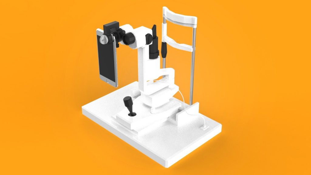
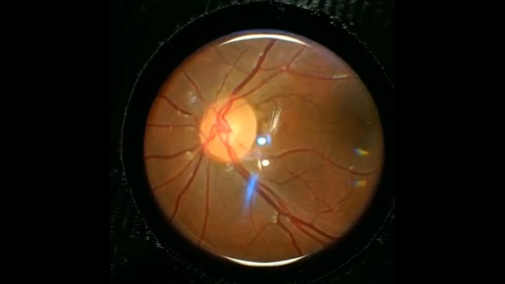
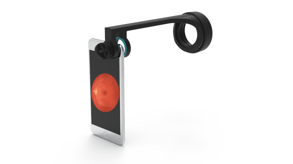
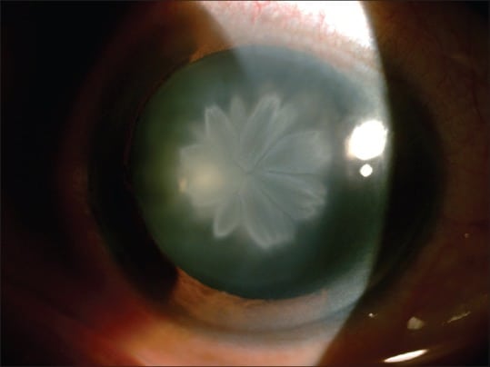
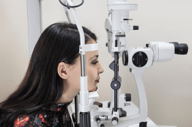
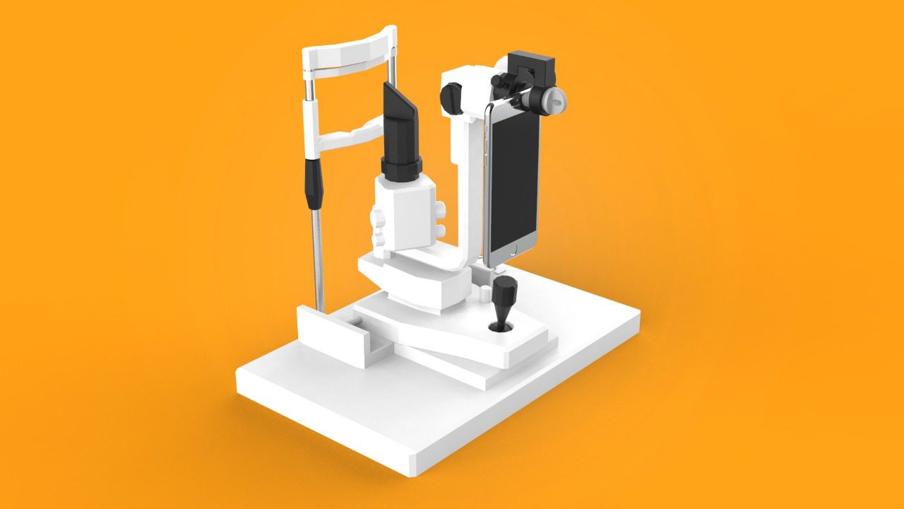
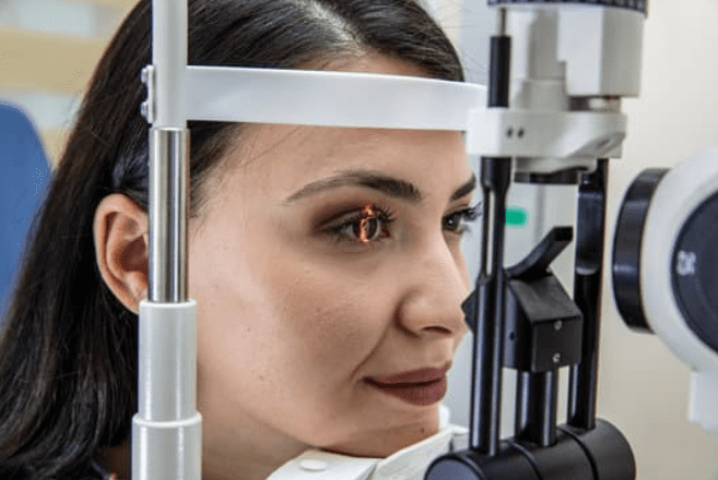
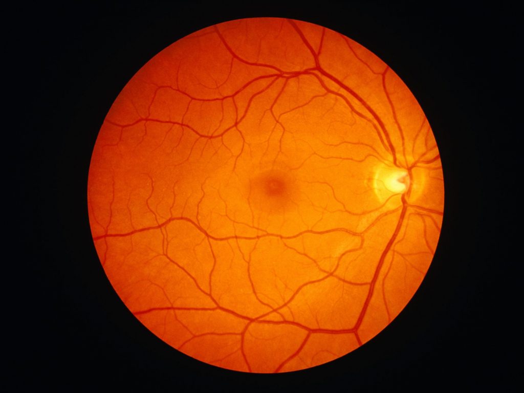
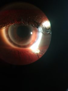
Smartphone slit-lamp photography is the new advancement in the field of science and technology in which the photographs of the desired slit-lamp finding can be taken with the smartphones by using the slit-lamp adapters. Uses of smartphone slit-lamp photography The smartphone slit-lamp photography allows for photo documentation of any ocular pathology. It has a full […]

Slit lamp exam purpose, uses, abnormal results & smartphone based slit lamp exam

Slit lamp examination

Slit lamp is the most important piece of equipment in the present-day ophthalmologist’s armamentarium. It is a routine method to examine the eyes for any abnormalities or diseases. A slit lamp is composed of three basic parts: Observation system a low-power binocular microscope mounted horizontally. Illumination system consists of a bright soured of light, a […]

Slit lamp examination : Biomicroscopy, types of illumination procedure of examination

Slit lamp examination

The slit lamp consists of two parts, an illumination system and a low-power binocular microscope mounted horizontally. The slit lamp examination is also known as biomicroscopy because it allows the in vivo study of living tissues of eye under the high magnification. Procedure of Slit lamp examination The examiner and patient are seated in either […]

Best 5 ways for retinal photography

Best 5 ways for retinal photography

Retinal Cameras Pros Digital images High Quality Different modes to view different retinal areas Good with hazy media Cons Very Expensive Needs DSLR Camera body to get digital images Not portable Needs skills and training Some types needs dilatation of pupil “Mydriatic types” Portable retinal devices Pros Portable Non mydriatic Digital images stored on the […]

Retinal imaging by smartphone | Retinal camera photography

Fundus examination is the most important part of eye care practice. The fundus photography is superior to the fundus examination as it allows for photo documentation of intraocular pathologies and for sharing of encrypted information with colleagues and patients. Traditional and portable fundus cameras are being used by ophthalmologists for this purpose. In recent times, […]

Subscribe & Get discount code