blog, Retina imaging
Retinal imaging by smartphone | Retinal camera photography
Oct
Fundus examination is the most important part of eye care practice. The fundus photography is superior to the fundus examination as it allows for photo documentation of intraocular pathologies and for sharing of encrypted information with colleagues and patients.
Traditional and portable fundus cameras are being used by ophthalmologists for this purpose. In recent times, smartphones have also been used for fundus imaging.
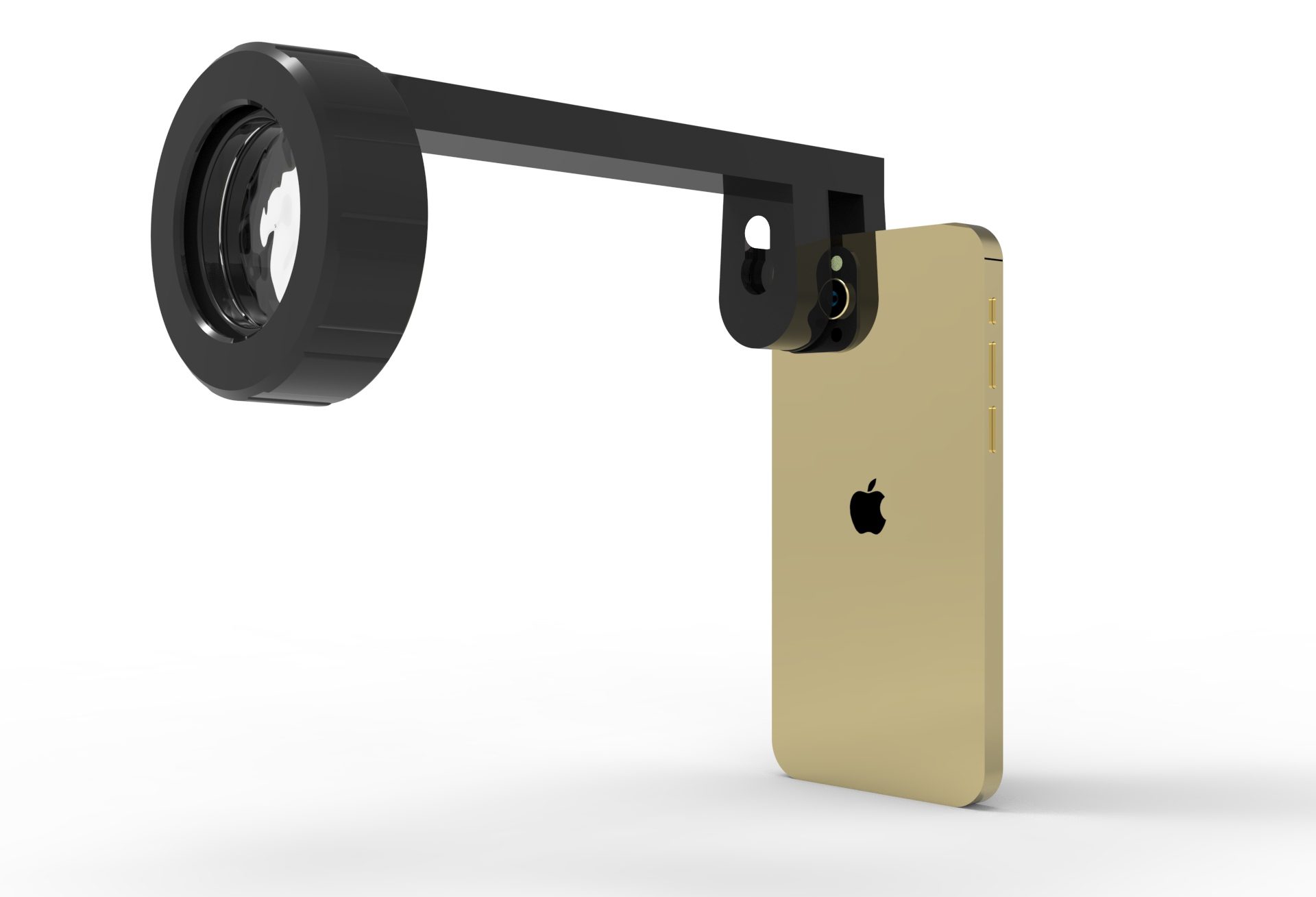
Recent technologies allow smartphone-based attachments and integrated lens adaptors to transform the smartphone into a portable fundus camera.
Why the smartphone fundus imaging is the best among all?
Traditional fundus cameras are expensive and have problems with convenience and portability. The demand to develop portable fundus cameras has increased the accessibility of retinal imaging, but most of these approaches rely on separate computers for viewing and transmission of fundus images.
In contrast, smartphones are readily available, relatively inexpensive, easy to operate. It has a fully embedded system capable of acquisition, storage and analysis of fundus images that can be directly transmitted from the phone via the wireless telecommunication system for remote evaluation.
Access to fundus photography is often limited by patient morbidity. This technique has been extremely helpful in the emergency department setting, during examinations under anesthesia, to document the fundus findings in infants and as a tool for fundus screening in camps, as it provides a cheaper and portable option for high quality fundus image acquisition for documentation and consultation.
Methods:
Smartphones with their high-resolution automatic focus cameras and light emitting diode (LED) light source has the potential to be used for fundus imaging. There are two methods:
* Smartphone Ophthalmoscope:
Smartphones are cost effective and can be used as cheap ophthalmoscope when used in conjunction with +20D lens.
* By slit lamp adapters:
To take slit lamp photos by placing the lens of your smartphone flush with the slit lamp ocular.
Smartphone ophthalmoscope for fundus photography
The smart phone camera can be used as a portable ophthalmoscope and use it for retinal photography when used in conjunction with a +20D condensing lens. The flash should be turned on to illuminate and view the fundus.
Steps:
- Dilate the patient pupil.
- Sit in front of the patient (20-30cm from the patient and at about the same level).
- Ask the patient to look at a far target.
- Set the smartphone camera on video mode.
- After darkening the room, turn the flash on and press the camera record button to record a continuous video.
- Hold the +20D lens in front of the patient’s eye (3-5cm from the patient cornea).
- Aim the light to the pupil and find the retinal glow.
Once you see the red reflex on your phone, you are basically in the right position. It takes practice to keep the camera, handheld lens, and patient pupil aligned.
While recording, move the camera and the lens to find a good focus and an image free of light reflections.
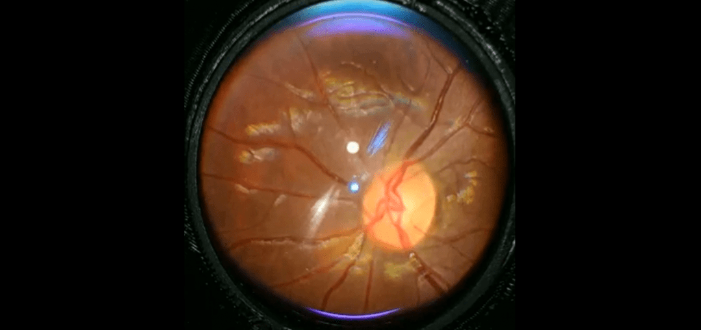
Adjust the handheld lens to and from the patient’s eye to see a clear retina image filling the entire lens area. Like an indirect ophthalmoscope view, this image is inverted.
To see the optic disc, ask the patient to look straight and fixate on a distant object.
To see the macula, ask the patient to look at the light.
To see the periphery, ask the patient to look right, left, up, up and left, up and right, down, down and left, down and right.
Play the video file back on your phone and pause at the frame of interest. Take a screenshot for each feature you wish to convey.
Use of Smart phones by slit lamp adapter:
Various slit-lamp adaptors have been created for ocular imaging with smartphones. These adapters attach the phone to the slit lamp and align the smartphone with the ocular of the slit lamp. The focusing and images are obtained through the smartphone’s camera apps and optical lens.
They consist of two parts – one part holds the smartphone securely, whereas the other part fixes onto the eyepiece of the slit lamp.
Most of these adaptors have been designed for anterior-segment imaging. For smartphone fundus imaging the use of these adaptors for slit-lamp has been successful however, the image quality has been poor due to difficulty with optics and illumination through the 78- or 90-D lenses on the slit lamp.
Steps and Tricks:
Flash is to be turned off for slit-lamp photographs, turned on for fundus photographs, Camera settings are to be set to maximum resolution for both photos and videos.
The patient should be illuminated well, from >1 diffuse light source with no areas of glare or reflections. Another smartphone in Torch/Flashlight mode can be used for this additional lighting if room lighting is not adequate.
Factors affecting the image quality
In smartphone retinal imaging, the image quality is very good but there are some factors which affect the image quality.
1. Glare :
The Pupil should be fully dilated in a minimal illuminated room to minimize glare during fundoscopy.
2. Light reflection :
Higher the Light reflections, Poorer the image quality. Try to eliminate unnecessary light reflections by adjusting the camera and lens positions. By holding the lens in slightly inclined position will eliminate maximum of light reflections.
3. Light intensity :
The patient comfort is the most important thing in examination, the intensity of most smartphone built in LEDs are extremely safe, the intensity is still excessive in dilating people. You may dim the light intensity by applying a semitransparent adhesive tape over the light source.
4. Media Opacities :
The fundus image quality is poor in the presence of ocular opacities e.g. Corneal opacities, Cataracts, vitreous opacities etc.
5. Field of view :
The field of view varies with the diopter strength of the handheld indirect ophthalmoscopy lens. Lower power lenses (such as +15 D or +20 D lenses) provide a smaller field of view with higher magnification.