
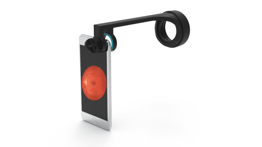
As ophthalmologists, we are constantly searching for new and innovative ways to improve our diagnostic capabilities, especially when it comes to detecting and managing glaucoma. One such technology that has recently emerged is the use of smartphone fundoscopes. A smartphone fundoscope is a device that attaches to the camera of a smartphone and allows […]
Category Archives: Retina imaging
29
Mar
Mar
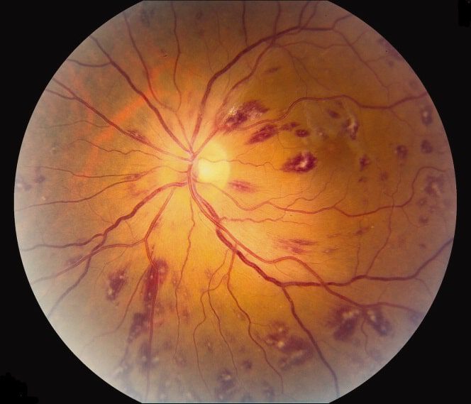
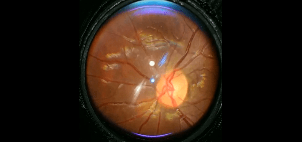
Roth spots case report A 46-year-old man presented with a four-week history of progressive bilateral visual loss. He also reported malaise, fever, anorexia, and night sweats, and had lost 14 kg in weight. He denied any history of exposure to HIV-related risk factors. Visual acuities at presentation were 6/36 in each eye with quiet anterior […]
26
Feb
Feb
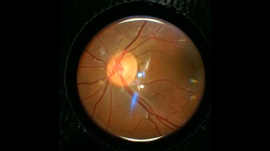
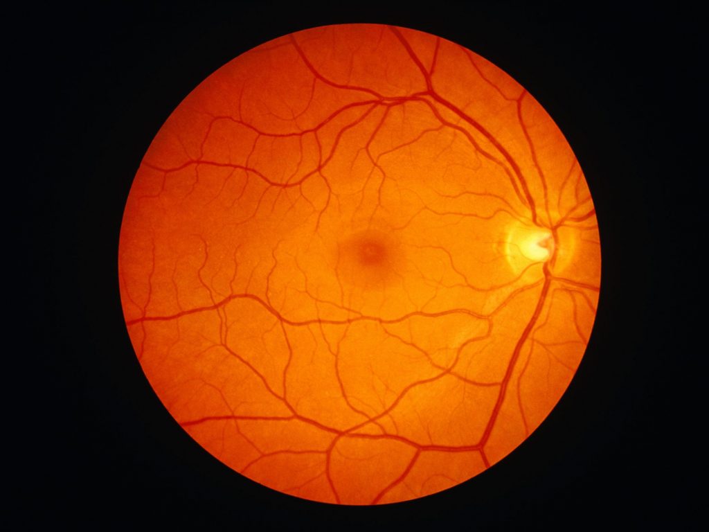
The most critical aspect of eye care practice is the Fundus examination. Fundus photography is superior to fundus analysis as it enables intraocular pathologies to be photo captured and encrypted information to be shared with colleagues and patients. Recent technologies allow smartphone-based attachments and integrated lens adaptors to transform the smartphone into a portable fundus […]
25
Feb
Feb
To get choroida fundus explorer, just follow these few steps: 1.Visit choroida.com and choose choroida innovative products. 2.Choose Fundus Explorer suitable with indirect fundus lenses. 3.From the lens list, choose between two options (with built-in 22D lens or without lens). 4.From the compatibility list, choose which lens you want your device to be compatible with […]
01
Dec
Dec
Retinal Cameras Pros Digital images High Quality Different modes to view different retinal areas Good with hazy media Cons Very Expensive Needs DSLR Camera body to get digital images Not portable Needs skills and training Some types needs dilatation of pupil “Mydriatic types” Portable retinal devices Pros Portable Non mydriatic Digital images stored on the […]
23
Oct
Oct
Fundus examination is the most important part of eye care practice. The fundus photography is superior to the fundus examination as it allows for photo documentation of intraocular pathologies and for sharing of encrypted information with colleagues and patients. Traditional and portable fundus cameras are being used by ophthalmologists for this purpose. In recent times, […]
- 1
- 2