blog
Why Do Many Ophthalmologists Avoid Routine Fundus Examination in Daily Practice?
Fundus examination remains a cornerstone in the diagnosis and follow-up of retinal and optic nerve diseases. Yet, in real-world clinical practice, many ophthalmologists do not perform it routinely during everyday clinic visits.
This hesitation is rarely due to a lack of awareness of its importance. Instead, it reflects a combination of practical, logistical, and workflow-related challenges that modern ophthalmologists face on a daily basis.
1. Time Constraints in High-Volume Clinics
In busy outpatient settings, efficiency is critical.
Routine fundus examination often requires additional steps:
- Pupil dilation
- Dark room conditions
- Prolonged chair time
In clinics with a high patient turnover, these steps can significantly disrupt workflow, leading some ophthalmologists to reserve fundus examination for selected cases rather than making it part of every visit.
2. Limited Access to Dedicated Fundus Imaging Systems
While tabletop fundus cameras provide high-quality images, they come with clear limitations:
- High acquisition and maintenance costs
- Space requirements
- Limited portability
As a result, many small or medium-sized clinics do not have in-house fundus imaging, forcing physicians to rely solely on indirect examination or to refer patients elsewhere for documentation.

3. Lack of Objective Documentation and Follow-Up
Contemporary ophthalmic practice increasingly depends on objective data:
- Image-based documentation
- Longitudinal comparison
- Multidisciplinary communication
Traditional fundus examination, although clinically valuable, does not inherently provide stored images. This makes follow-up comparisons difficult and limits the ability to clearly explain findings to patients, trainees, or referring physicians.
4. Dependence on Examiner Experience
Accurate fundus examination requires:
- Technical skill
- Experience in image interpretation
- Consistent examination conditions
Inter-observer variability remains a challenge, particularly in non-specialized settings or training environments. Subtle changes may be overlooked, especially during brief or high-pressure consultations.
5. Patient Cooperation and Comfort
In daily practice, not all patients are ideal candidates for conventional fundus examination:
- Pediatric patients
- Elderly individuals
- Patients with photophobia or limited cooperation
These factors can reduce examination quality and discourage routine use, especially when alternative, faster assessment methods are available.
6. The Gap Between Clinical Importance and Practical Feasibility
Despite its undeniable diagnostic value, fundus examination often competes with the realities of modern ophthalmic practice. The gap lies not in understanding its importance, but in finding solutions that align with:
- Clinic workflow
- Time efficiency
- Documentation needs
- Portability
Bridging this gap is essential for improving routine retinal assessment without increasing the clinical burden on ophthalmologists.
7. Absence of Immediate Visual Feedback
Unlike anterior segment imaging, traditional fundus examination does not provide immediate visual confirmation that can be reviewed after the encounter.
Once the examination is completed, the findings rely entirely on the examiner’s real-time observation and written description.
This limitation:
- Reduces the ability to re-evaluate subtle findings
- Makes peer discussion more difficult
- Limits the educational value during case reviews
In an era where visual documentation is central to clinical decision-making, this lack of instant visual feedback can discourage routine fundus assessment.
8. Challenges in Patient Education and Engagement
Patients increasingly expect to understand their condition visually.
Explaining retinal findings without images can be difficult, particularly for chronic diseases such as diabetic retinopathy or macular degeneration.

Without visual aids:
- Patient understanding may be limited
- Compliance with treatment and follow-up can be reduced
- Trust-building opportunities may be missed
This communication gap can make fundus examination feel less impactful during routine visits.
9. Limited Educational Value for Trainees in Busy Clinics
In teaching environments, fundus examination is a valuable learning opportunity. However, in high-volume clinics:
- There is limited time for supervised examination
- Trainees often rely on verbal descriptions rather than shared visual findings
- Subtle pathology may go unnoticed without image reference
This reduces the consistency of training and may discourage routine examination in favor of faster, less instructive workflows.
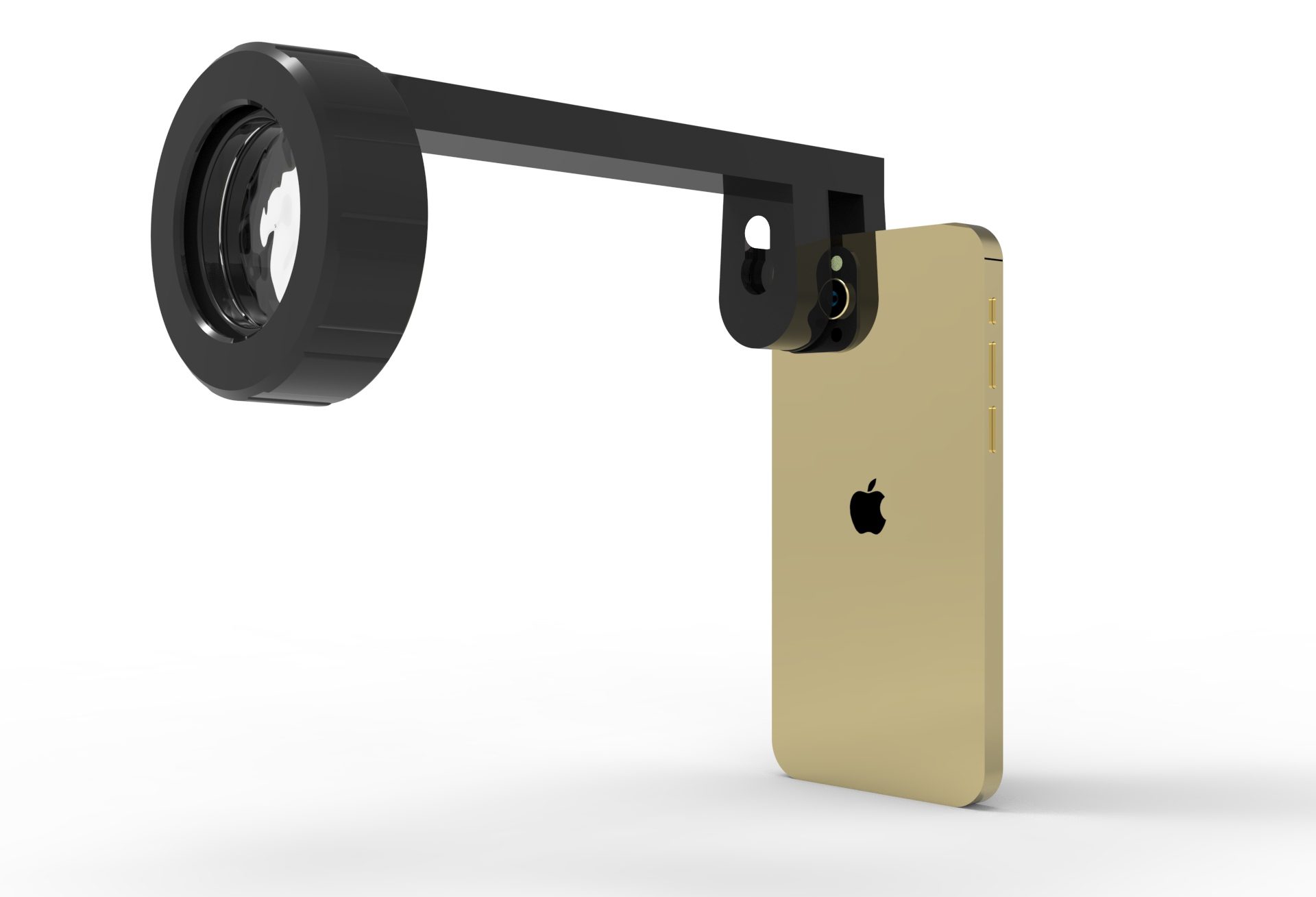
Smartphone Fundoscopy as a Practical Solution in Daily Practice
Recent advances in smartphone fundoscopy have helped bridge many of the gaps associated with traditional fundus examination. Devices such as the Fundus Explorer Pro illustrate how portable imaging solutions can be integrated into routine clinical workflows without adding complexity.

By enabling rapid fundus imaging using a smartphone, this approach helps address several of the previously mentioned challenges:
- Time efficiency, allowing quick documentation without disrupting clinic flow
- Objective image documentation for follow-up, comparison, and case discussion
- Improved patient communication, as retinal findings can be visualized and explained directly
- Enhanced educational value, especially for trainees, through shared and reviewable images
Rather than replacing clinical examination, smartphone fundoscopy serves as a practical extension—making fundus assessment more accessible, repeatable, and easier to incorporate into everyday ophthalmic practice.
References
American Academy of Ophthalmology (AAO)
Funduscopic Examination.
American Academy of Ophthalmology Clinical Statements.
Russo A, Morescalchi F, Costagliola C, et al.
Comparison of smartphone ophthalmoscopy with slit-lamp biomicroscopy for retinal evaluation.
American Journal of Ophthalmology. 2015;159(2):360–364.
Wintergerst MWM, Brinkmann CK, Holz FG, Finger RP.
Underserved populations and the role of mobile ophthalmic imaging.
Survey of Ophthalmology. 2020;65(5):593–602.
Bastawrous A, Giardini ME, Bolster NM, et al.
Clinical validation of a smartphone-based adapter for retinal imaging.
Eye (Lond). 2016;30(8):1162–1168.
Rajalakshmi R, Subashini R, Anjana RM, Mohan V.
Automated diabetic retinopathy detection in smartphone-based fundus photography.
Investigative Ophthalmology & Visual Science. 2018;59(4):1422–1428.