blog
Retinopathy of Prematurity Screening with a Smartphone: A Practical Guide
Retinopathy of prematurity is one of the leading preventable causes of childhood blindness worldwide, and its burden is rising as neonatal survival improves in low- and middle-income regions.
The cruelty of ROP is its timing: the window in which treatment prevents lifelong blindness is narrow, often just days, and it closes whether or not a trained examiner is available.
In much of the world, the rate-limiting step is not knowledge of how to treat ROP — it is getting an expert pair of eyes to the right baby at the right time.
This is where imaging changes the arithmetic. Retinopathy of prematurity screening supported by retinal imaging lets a non-specialist capture the infant’s fundus and a specialist grade it remotely — and increasingly, that image can be captured on a smartphone.
This guide covers who needs screening and when, how smartphone-assisted ROP imaging fits, the technique, and the essential safety caveats unique to neonatal examination.
 ROP by the numbers
|
A leading preventable cause of
childhood blindness
|
Treatment window once the threshold is reached
often days
|
|
Typical screening trigger (varies)
≤30–31 wks / ≤1500 g
|
Telemedicine ROP grading (studies)
high concordance
|
Sources: WHO/IAPB childhood blindness reports; national ROP screening guidelines; telemedicine ROP validation studies. Always follow your local guidelines for thresholds.
Why is ROP screening capacity the real bottleneck
ROP screening is labour-intensive and specialist-dependent. It requires repeated, time-critical examinations of fragile infants by someone trained to recognise zone, stage and plus disease — a skill set concentrated in a small number of ophthalmologists who cannot be physically present in every neonatal unit on the day each baby reaches threshold. As survival of very preterm infants rises, demand outstrips that workforce, and babies in smaller or remote units are the ones who fall through the gap.
Imaging-based, telemedicine-supported screening directly attacks this bottleneck. If a trained nurse or non-specialist can capture adequate retinal images at the cot-side and transmit them, one specialist can grade infants across many units without travelling to each — multiplying scarce expertise rather than rationing it.
What the evidence says about image-based ROP screening
Telemedicine for ROP, using wide-field digital retinal images graded remotely, has a substantial evidence base and is endorsed within several national screening frameworks as a valid model when implemented with appropriate quality control.
Studies of remote grading report high concordance with bedside indirect ophthalmoscopy for clinically significant disease. The principle is well established; the practical barrier has been the cost and availability of dedicated wide-field neonatal imaging systems.
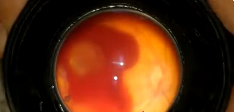
Smartphone-assisted imaging lowers that barrier. While dedicated wide-field contact systems remain the reference for formal ROP imaging programmes, smartphone fundoscopy through a condensing lens has been described as a low-cost adjunct for documentation, education and screening support — particularly in settings where a dedicated system is simply not available, and where the alternative is no image at all.
Where a smartphone fits — and the design that matters
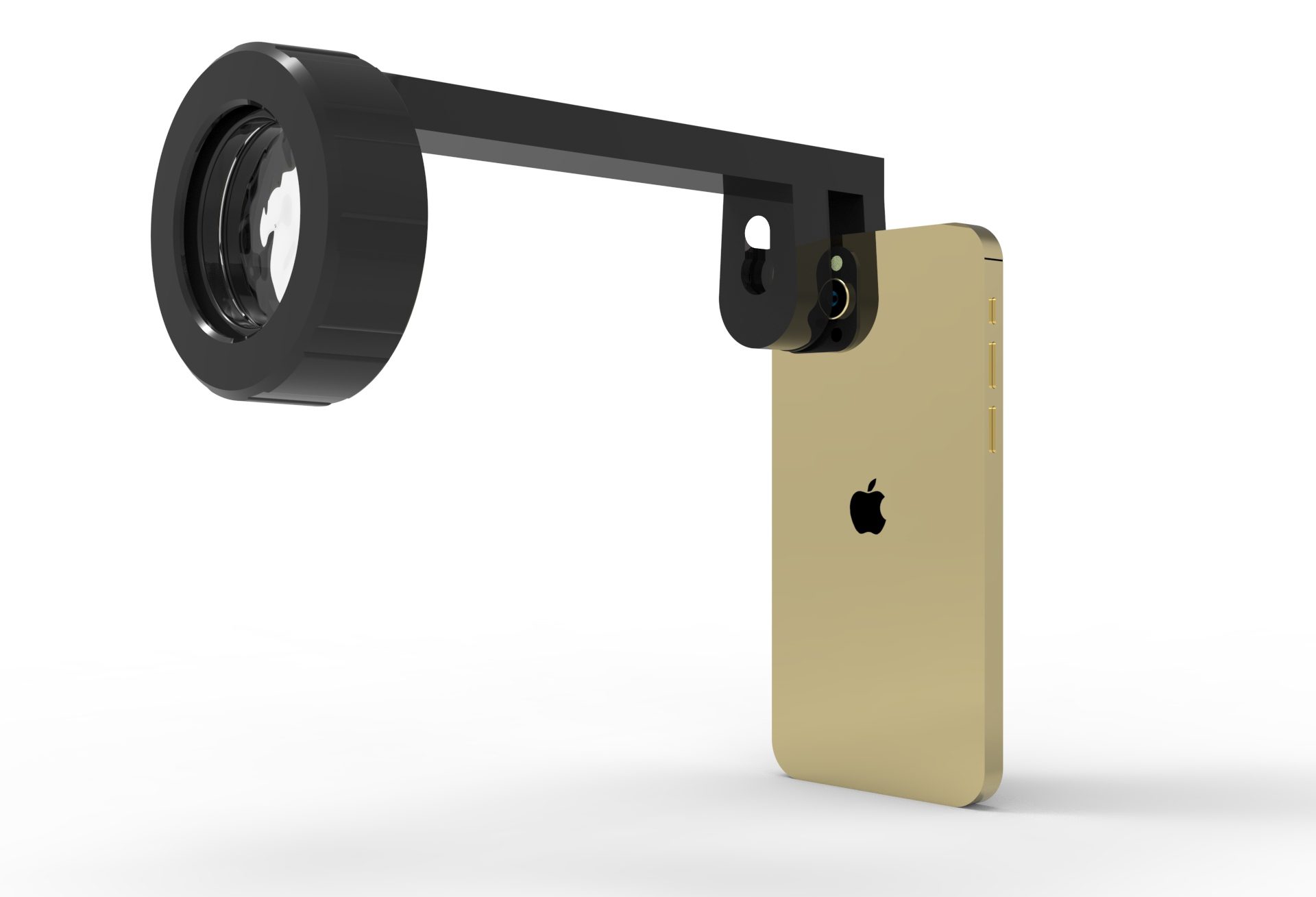
For neonatal imaging, two device qualities matter even more than usual: a light source that is effective but not reliant on a harsh phone flash, and compatibility with a condensing lens to deliver an indirect view.
This is the clinical justification for the design of the Fundus Explorer Pro: a dedicated 150-lumen LED independent of the phone flash, and a 22D indirect lens giving a field of view beyond 60°, on any smartphone — putting documentation and tele-grading support within reach of units that could never fund a dedicated wide-field system.
It is essential to be clear about the scope: a smartphone device is an adjunct and documentation tool.
It supports screening, teaching and tele-consultation; it does not replace examination by an ophthalmologist trained in ROP, nor the wide-field contact systems used in formal imaging programmes.
Used within that scope, it extends reach; pushed beyond it, it risks false reassurance.
A practical, safety-first workflow
- 1Follow your local screening guideline.Gestational-age/birth-weight thresholds and timing differ between countries — the guideline, not the device, sets who and when.
- 2Prepare the infant safely.Dilation, swaddling, monitoring and the presence of neonatal staff per protocol; minimise examination time and handling.
- 3Capture systematically.Disc and posterior pole first, then sweep the zones; use video to maximise the chance of usable frames in a brief, tolerated window.
- 4Document zone, stage and plus signs.Note vascular dilation/tortuosity (plus disease) and the most posterior zone of disease — these drive urgency.
- 5Transmit for specialist grading.Send labelled, de-identified images promptly; escalate any suspicion of plus or posterior disease immediately rather than waiting.
What demands urgent escalation
- ▲
Plus disease — venous dilation and arteriolar tortuosity at the posterior pole.
- ▲
Any disease in zone I, or aggressive posterior ROP — highest urgency.
- ▲
Stage 3 with plus, or rapid progression — or ungradable images — repeat or refer; never assume normal.
- ▼
Mature, fully vascularised retina — meeting your guideline’s exit criteria.
“In ROP, distance is the disease’s ally. An image that a non-specialist can capture and a specialist can grade from afar takes that ally away.”
 Is the Fundus Explorer Pro right for ROP support?
|
Best suited for
|
Keep in mind
|
The bottom line
ROP blinds children not because we cannot treat it, but because the right expertise too often cannot reach the right baby inside a window measured in days.
Image-based, telemedicine-supported screening shortens that distance, and smartphone-assisted imaging puts the entry point within reach of units that could never fund a dedicated system.
As a documentation and tele-grading adjunct — never a replacement for ROP-trained examination — a device like the Fundus Explorer Pro helps turn scarce specialist time into wider reach, and a closing window into a caught diagnosis.
References: WHO/IAPB reports on childhood blindness and ROP; national ROP screening guidelines (e.g., AAP/AAPOS and equivalents); telemedicine ROP validation literature on remote digital image grading; descriptions of smartphone-assisted neonatal fundus imaging. Always follow your local screening guideline for thresholds and timing.