blog, Retina imaging
Science behind smartphone fundoscopy
Jul
The advent of smartphones has ushered in a new era of accessibility and convenience in various aspects of our lives, including healthcare.
One remarkable application of smartphone technology in the medical field is smartphone fundoscopy, which enables healthcare professionals and even individuals to capture high-resolution images of the eye’s fundus using just a smartphone.
This innovative approach has the potential to revolutionize eye health by providing a cost-effective, portable, and easy-to-use solution for diagnosing and monitoring various eye conditions. In this article, we will delve into the science behind smartphone fundoscopy and explore its implications for eye care.
Understanding Fundoscopy
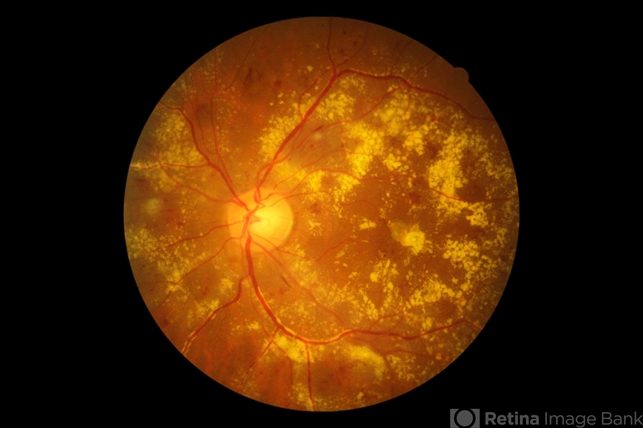
Fundoscopy, also known as ophthalmoscopy, is a technique used by ophthalmologists and optometrists to examine the fundus of the eye. The fundus refers to the inner lining of the eye, which includes the retina, optic disc, blood vessels, and other structures crucial for vision.
By examining the fundus, healthcare professionals can detect signs of various eye diseases, such as diabetic retinopathy, glaucoma, and macular degeneration.
Traditionally, fundoscopy requires specialized equipment like a direct ophthalmoscope or a slit-lamp biomicroscope. These tools are costly, bulky, and require expertise to operate, making them inaccessible in certain settings and underserved regions.
Smartphone fundoscopy, on the other hand, leverages the powerful imaging capabilities of smartphones and addresses these limitations.
The Science Behind Smartphone Fundoscopy
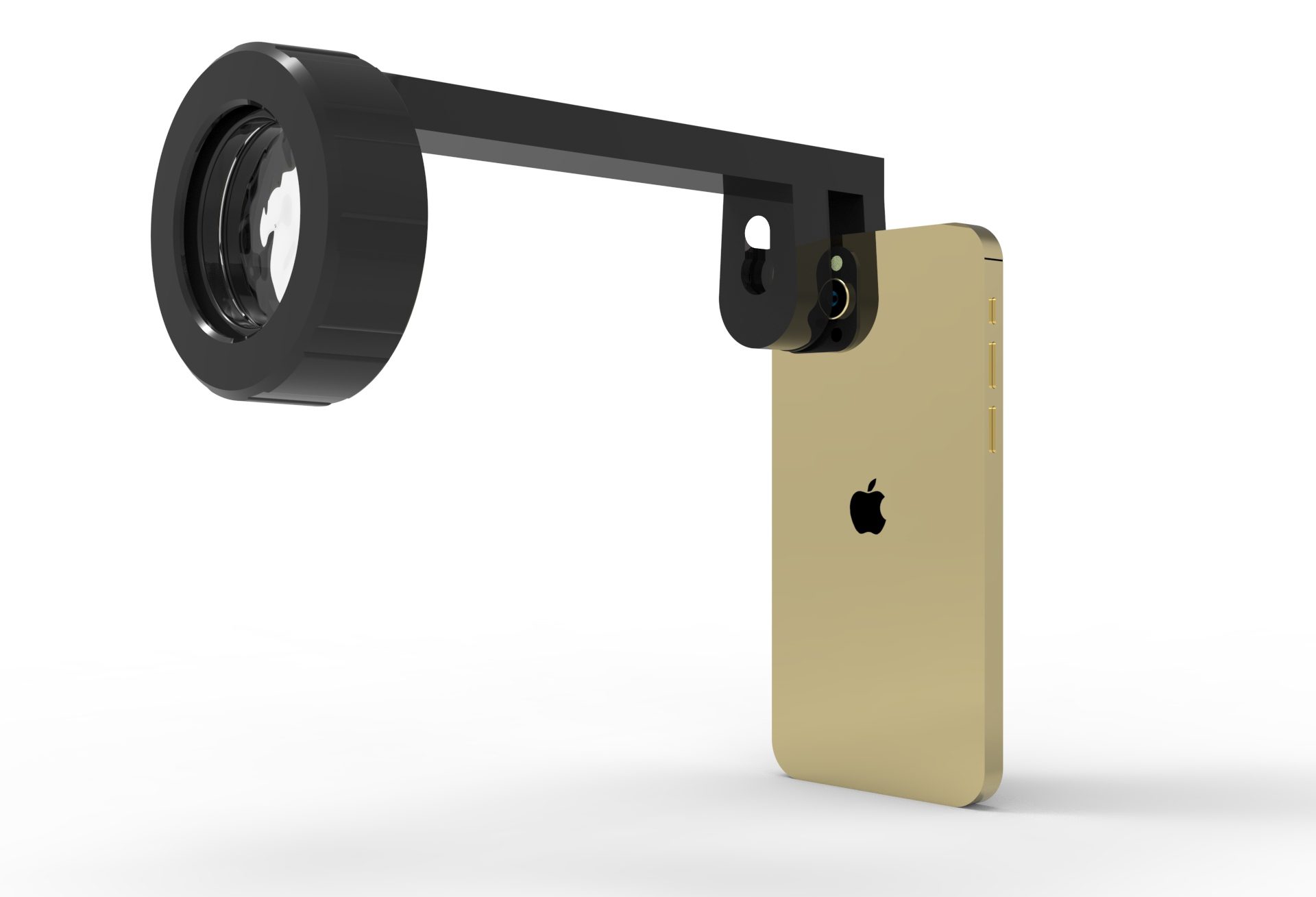
Smartphone fundoscopy relies on the integration of three key components: the smartphone’s camera, a light source, and an adaptor. Let’s explore how these components work together to capture detailed images of the fundus:
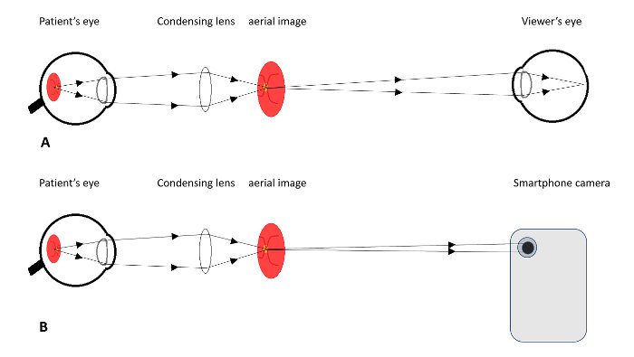
Camera: Modern smartphones are equipped with high-resolution cameras capable of capturing detailed images. These cameras utilize advanced imaging sensors and lenses to produce clear and focused pictures. For fundoscopy, the smartphone’s camera is aligned with the eye’s pupil to capture the fundus image.
Light Source: Adequate illumination is crucial for fundoscopy. Traditional fundoscopes use a built-in light source, but smartphones utilize the camera’s flash or an external light source. The light is projected into the eye through the pupil, illuminating the fundus for better visibility and image capture.
Adaptor: To align the smartphone’s camera with the eye’s pupil, various adaptors or attachments are available in the market. These adaptors are designed to hold the smartphone securely and ensure proper alignment for optimal image quality. Some adaptors incorporate lenses to enhance the field of view or compensate for refractive errors.
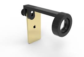
Image Enhancement and Analysis
Once the fundus images are captured using a smartphone, they can be further enhanced and analyzed using specialized software applications. These applications employ image processing algorithms to improve image clarity, adjust contrast and brightness, and remove artifacts or noise.
Some advanced applications even incorporate artificial intelligence algorithms for the automatic detection of abnormalities, assisting healthcare professionals in diagnosing and monitoring eye conditions more efficiently.
Benefits
The science behind smartphone fundoscopy offers several benefits that have the potential to revolutionize eye health:
Accessibility: Smartphone fundoscopy democratizes access to eye care by bringing it to remote or underserved areas where traditional equipment is scarce. It allows for screening and diagnosis to be performed outside traditional clinical settings, increasing the reach of eye care services.
Cost-effectiveness: Compared to traditional fundoscopy equipment, smartphones are affordable and widely available. Smartphone fundoscopy significantly reduces the financial burden associated with acquiring and maintaining specialized equipment, making it a cost-effective alternative.
Portability and Convenience: Smartphones are portable and easily transportable, enabling eye examinations to be conducted in various settings. This portability is especially beneficial for emergency care, telemedicine, or mobile healthcare units.
Despite its numerous advantages, smartphone fundoscopy does have some limitations. It relies on the user’s ability to properly align the camera, which may lead to suboptimal image quality if not done correctly. Additionally, smartphone fundoscopy may not replace the need for traditional fundoscopy in complex cases that require a comprehensive examination by a trained eye care professional.
Conclusion
The science behind smartphone fundoscopy holds immense promise for transforming eye care. By leveraging the power of smartphones, this innovative technique makes fundus imaging more accessible, cost-effective, and portable.

Smartphone fundoscopy has the potential to extend the reach of eye care services, improve the early detection of eye diseases, and facilitate remote monitoring. As technology continues to advance, smartphone fundoscopy is expected to play an increasingly significant role in promoting eye health worldwide.