
No products in the cart.
blog, Retina imaging
Fundus photography Diagnostic Power: a comprehensive review
21
Mar
Mar
Fundus photography stands at the forefront of modern ophthalmology, offering clinicians an unparalleled window into the inner workings of the eye.

This sophisticated imaging technique has revolutionized the field, enabling early detection, precise monitoring, and effective management of a wide array of ocular conditions.
From diabetic retinopathy to age-related macular degeneration, fundus photography serves as a cornerstone in the diagnostic armamentarium of ophthalmologists worldwide.
Evolution of Fundus Photography
The evolution of fundus photography can be traced back to the late 19th century, with the pioneering work of ophthalmologists such as Wilhelm Friedrich Ostwald and Carl Pulfrich.
Ostwald’s adaptation of the ophthalmoscope for photography laid the groundwork for capturing detailed images of the ocular fundus.
Over the ensuing decades, advancements in optics, illumination, and imaging technology propelled fundus photography into the digital era.
Fundamentals of Fundus Photography
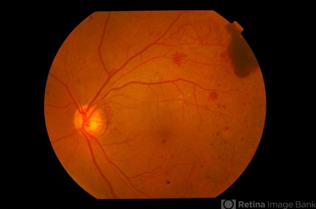
At its core, fundus photography involves capturing high-resolution images of the posterior segment of the eye, including the retina, optic nerve head, and macula.
The process typically begins with pupil dilation to optimize visualization of the fundus. A specialized camera equipped with filters, focusing mechanisms, and illumination sources is then used to capture images of the ocular fundus.
Clinical Applications
Diabetic Retinopathy:
Diabetic retinopathy remains a leading cause of vision loss globally, emphasizing the critical role of early detection and intervention.
Fundus photography plays a pivotal role in screening diabetic patients for retinopathy, enabling timely referral for further evaluation and treatment.
High-quality fundus images facilitate the assessment of microaneurysms, hemorrhages, exudates, and neovascularization, guiding treatment decisions and monitoring disease progression.
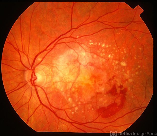
Age-Related Macular Degeneration (AMD):
AMD represents another major public health concern, particularly in aging populations. Fundus photography serves as a cornerstone in the diagnosis and management of AMD, allowing clinicians to visualize drusen, pigmentary changes, geographic atrophy, and choroidal neovascularization.
Sequential fundus images aid in tracking disease progression, evaluating treatment efficacy, and informing therapeutic strategies such as anti-VEGF therapy or photodynamic therapy.
Glaucoma:
While glaucoma primarily affects the optic nerve, its impact on the retinal nerve fiber layer (RNFL) underscores the utility of fundus photography in its diagnosis and monitoring.
Optic disc morphology, neuroretinal rim integrity, and RNFL thickness can be meticulously evaluated using fundus images, complementing other diagnostic modalities such as optical coherence tomography (OCT).
Serial fundus photography enables longitudinal assessment of glaucomatous changes, guiding treatment decisions and optimizing patient care.
Retinal Vascular Diseases:
Fundus photography plays a crucial role in the diagnosis and management of various retinal vascular diseases, including hypertensive retinopathy, central retinal vein occlusion (CRVO), and branch retinal vein occlusion (BRVO).
Characteristic findings such as arteriolar narrowing, arteriovenous nicking, cotton-wool spots, and retinal hemorrhages are meticulously documented through fundus imaging, aiding in risk stratification, prognostication, and treatment planning.
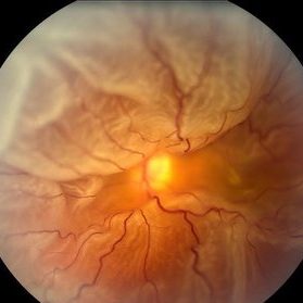
Retinal Detachment:
In cases of retinal detachment, fundus photography serves as a valuable tool for preoperative assessment and surgical planning.
Detailed fundus images help identify the extent and location of retinal breaks, delineate associated proliferative vitreoretinopathy (PVR), and document associated findings such as choroidal detachment or subretinal fluid.
Postoperatively, fundus photography facilitates the evaluation of surgical outcomes and the detection of recurrent detachments or complications.
Technological Advancements
Widefield Imaging:
Recent advancements in imaging technology have expanded the utility of fundus photography, with the advent of widefield imaging systems.
These systems offer panoramic views of the fundus, capturing peripheral retinal pathology that may be missed with conventional imaging techniques.
Widefield fundus photography is particularly valuable in conditions such as peripheral retinal degenerations, retinal tears, and peripheral vascular abnormalities, enhancing diagnostic accuracy and guiding treatment decisions.
Autofluorescence Imaging:
Autofluorescence imaging represents another innovative application of fundus photography, leveraging the intrinsic fluorescence properties of ocular tissues.
This non-invasive imaging modality provides insights into retinal pigment epithelial (RPE) function and the accumulation of lipofuscin, aiding in the diagnosis and monitoring of various retinal disorders.
Autofluorescence abnormalities observed on fundus images can serve as biomarkers for disease activity and progression in conditions such as geographic atrophy and inherited retinal dystrophies.
Angiography:
Fluorescein angiography (FA) and indocyanine green angiography (ICGA) complement fundus photography by providing dynamic visualization of retinal and choroidal circulation, respectively.
These angiographic techniques facilitate the assessment of retinal perfusion, vascular leakage, and neovascularization in conditions such as diabetic retinopathy, AMD, and retinal vascular occlusions.
Integration of angiographic findings with fundus photography enhances diagnostic precision and informs therapeutic interventions such as laser photocoagulation or anti-angiogenic therapy.
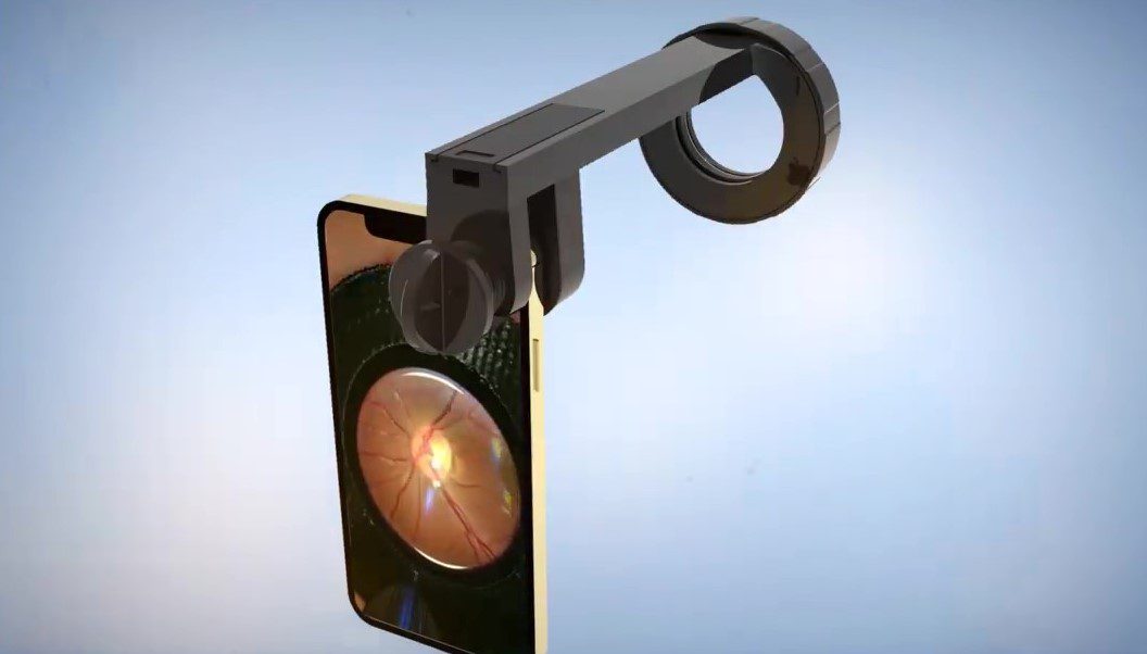
Smartphone Fundoscopy:
With the ability to capture high-quality images of the retina using a smartphone camera, this technology has revolutionized the field, making eye examinations more accessible and convenient than ever before providing accessible, cost-effective screening and diagnostic capabilities.
It has the potential to improve early detection, facilitate remote consultations, empower patients, and enhance medical education.
Fundus Explore Pro is an innovative device that represents a significant advancement in fundus imaging technology.
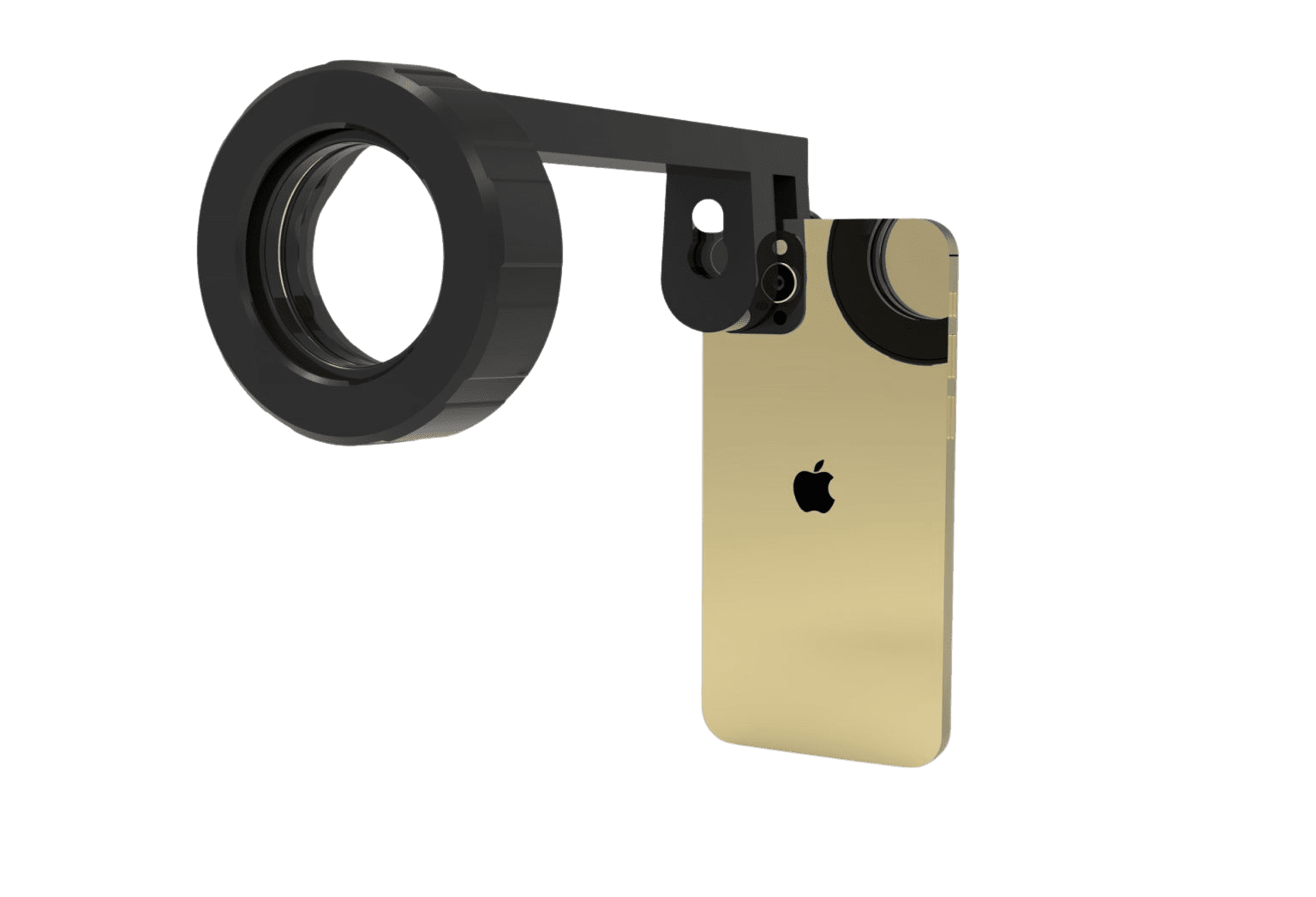
Unlike traditional fundus cameras, which are bulky and expensive, Fundus Explorer Pro is a compact attachment that can be easily affixed to a smartphone’s camera lens.
Challenges and Future Directions
Despite its undeniable utility, fundus photography faces challenges related to accessibility, cost, and interpretability, particularly in resource-limited settings.
Addressing these challenges requires ongoing innovation in imaging technology, workflow optimization, and training programs for healthcare providers.
Furthermore, the integration of artificial intelligence (AI) and machine learning algorithms holds promise for automating image analysis, facilitating early disease detection, and personalized treatment planning.
Looking ahead, the future of fundus photography in ophthalmology is poised for remarkable advancements, fueled by interdisciplinary collaborations, technological innovation, and a shared commitment to optimizing patient care.
As we continue to unravel the complexities of ocular pathology, fundus photography will undoubtedly remain a cornerstone in the diagnostic arsenal of ophthalmologists worldwide, empowering clinicians to preserve and restore vision for generations to come.
You may be interested in reading:
- Fundus examination: An In-Depth Look and Innovative Solution
- Exploring Fundus Cameras and Smartphone Integration in Fundus Examination