blog, Retina imaging
Exploring Fundus Cameras and Smartphone Integration in Fundus Examination
Mar
Human eye is a complex organ with various components contributing to the vision. The fundus, the interior surface of the eye opposite the lens, is particularly important for assessing eye health.
Fundus examination plays a crucial role in diagnosing and monitoring various ocular conditions, such as diabetic retinopathy, glaucoma, macular degeneration, and hypertension-related eye disorders.
Fundus cameras are essential tools in this regard, providing high-resolution images of the fundus for analysis by healthcare professionals.
In recent years, the integration of smartphone fundoscopy through specialized attachments has emerged as a promising approach to widen access to fundus examination, particularly in resource-limited settings.
Fundus Cameras: Imaging Technique and Applications
Fundus cameras are sophisticated imaging devices specifically designed to capture detailed images of the fundus.
They utilize various imaging techniques to produce high-quality photographs that aid in the diagnosis and management of ocular diseases. The imaging process typically involves the following steps:
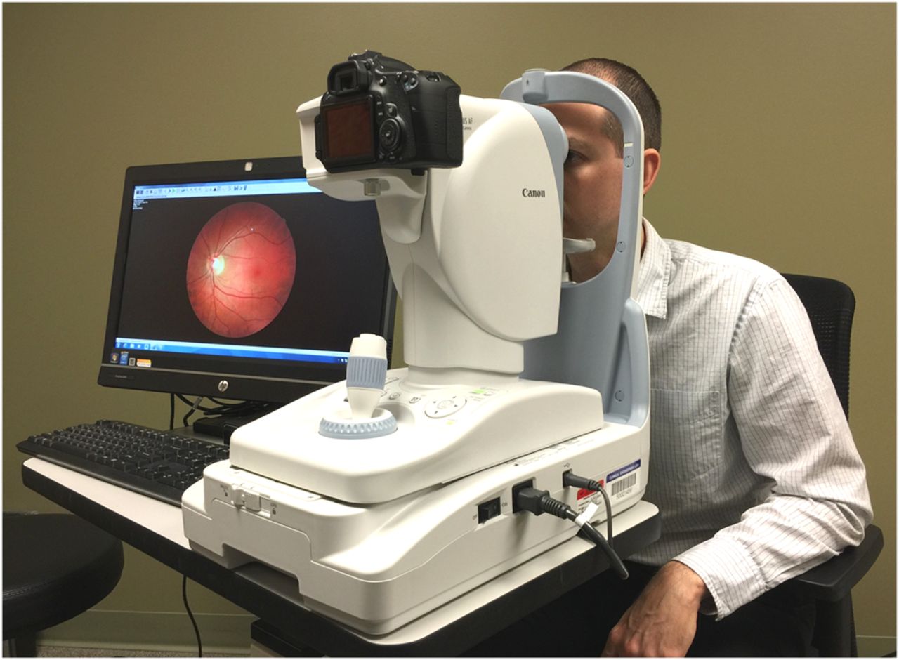
Pupil Dilation: Before capturing fundus images, the patient’s pupils are dilated to ensure a wide view of the fundus.
Alignment and Focusing: The patient is positioned correctly, and the camera is aligned to capture images of the desired area of the fundus. Precise focusing is crucial to obtaining sharp and detailed images.
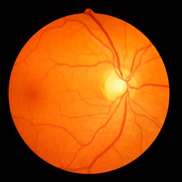
Image Capture: Once the camera is aligned and focused, it captures digital images of the fundus. Fundus cameras employ various illumination techniques to illuminate the fundus and enhance visibility.
Image Processing and Analysis: The captured images are then processed and analyzed using specialized software.
This software allows healthcare professionals to zoom in on specific areas of interest, adjust contrast and brightness, and perform measurements for diagnostic purposes.
Fundus cameras find wide-ranging applications in ophthalmology, optometry, and general medicine. They are indispensable tools for screening, diagnosing, and monitoring ocular diseases.
Early detection of conditions such as diabetic retinopathy is crucial for timely intervention and prevention of vision loss.
Smartphone Integration in Fundus Examination
While traditional fundus cameras offer high-quality imaging, they are often expensive and require specialized training to operate.
Moreover, their bulky size and reliance on external power sources limit their portability and accessibility, especially in remote or underserved areas.
Recognizing these limitations, researchers have explored alternative approaches to fundus imaging, leveraging the ubiquity and technological capabilities of smartphones.
Special Attachments for Smartphone Fundus Imaging
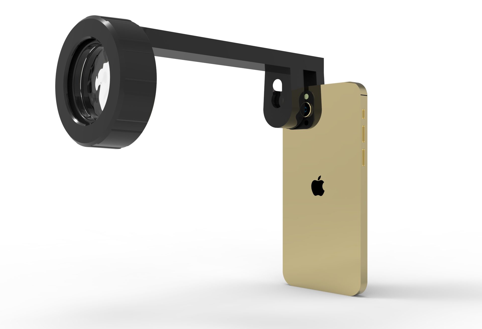
Specialized attachments have been developed to transform smartphones into portable fundus cameras like Fundus Explorer Pro device.
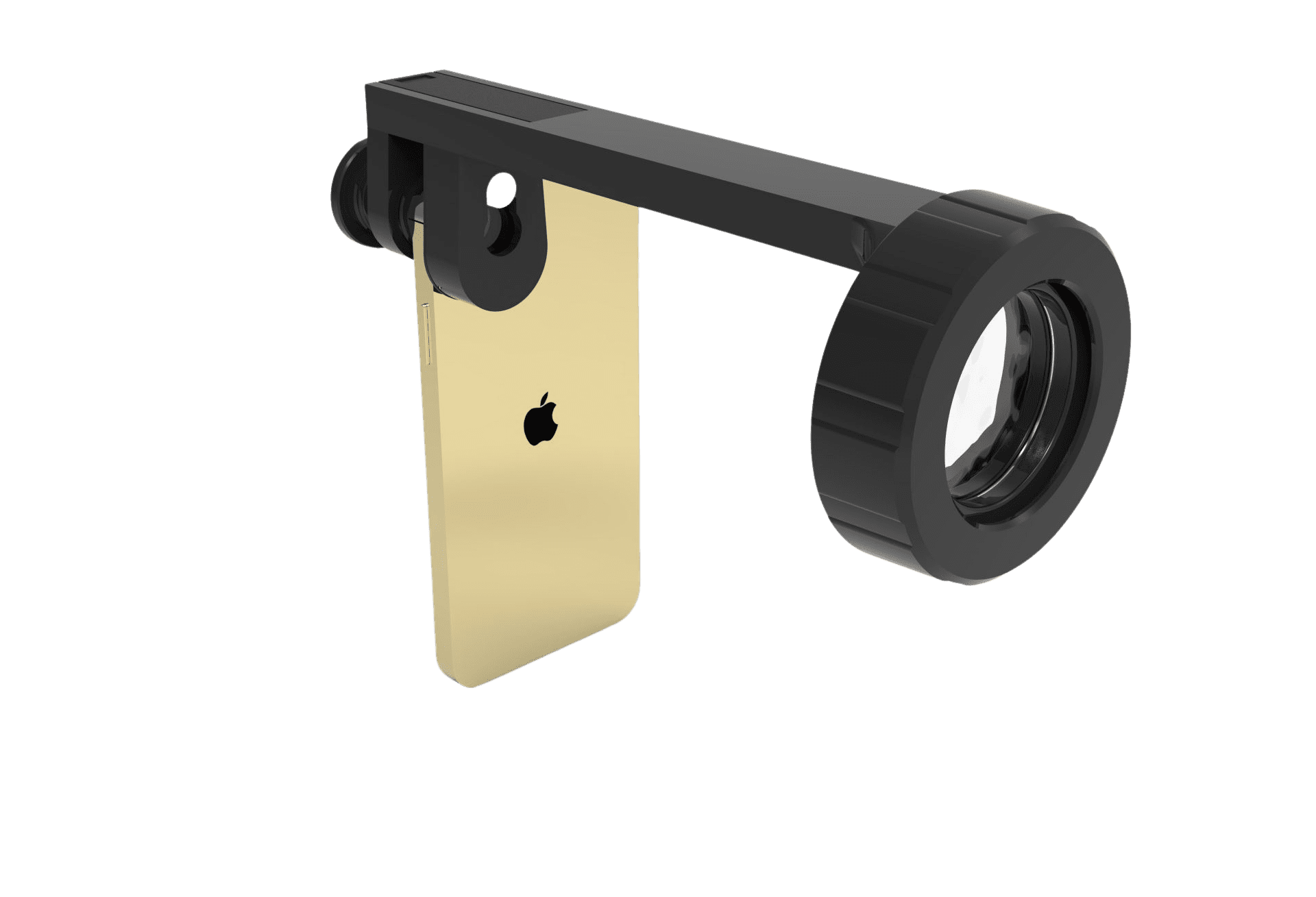
These attachments typically consist of a lens system and an illumination source that attaches to the smartphone’s camera module.
Imaging Technique with Smartphone Attachments
The imaging technique with smartphone attachments follows a similar process to traditional fundus photography but with some variations:
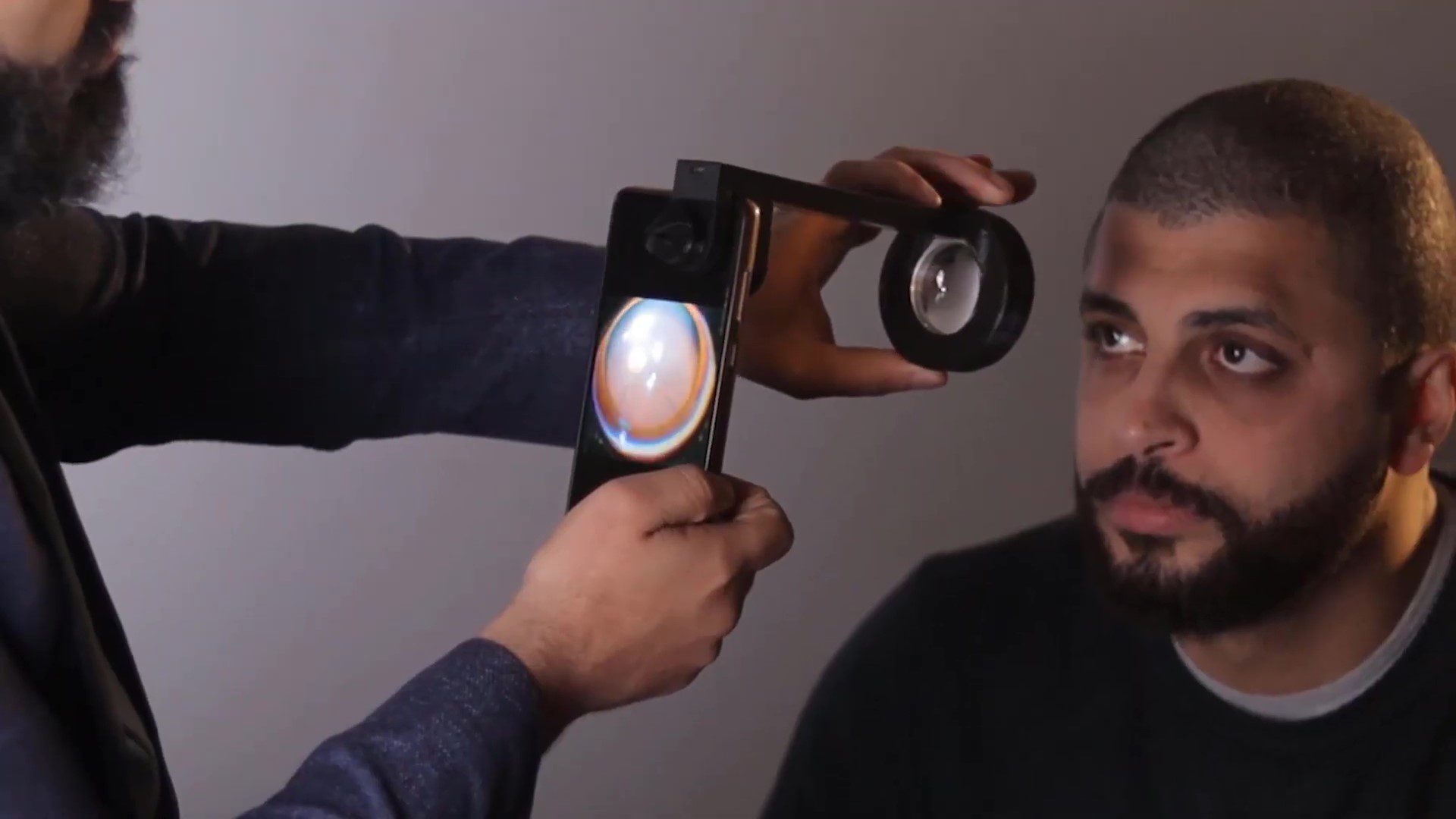
Attachment Setup: The smartphone attachment is securely mounted onto the smartphone’s camera module, aligning it with the lens and ensuring proper illumination.
Patient Preparation: Similar to traditional fundus examination, the patient’s pupils may need to be dilated using eye drops to obtain clear images of the fundus.
Alignment and Focusing: The smartphone is aligned with the patient’s eye, and the attachment is focused to capture images of the fundus.
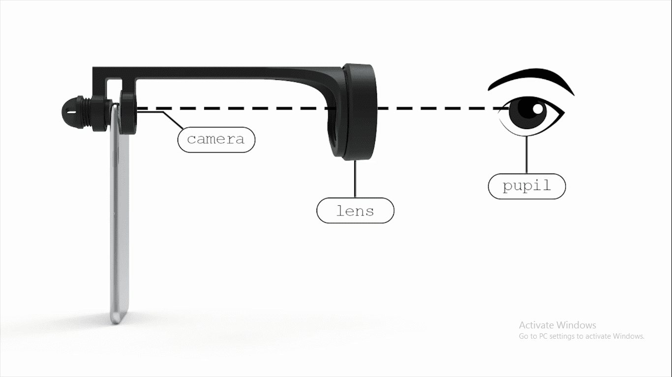
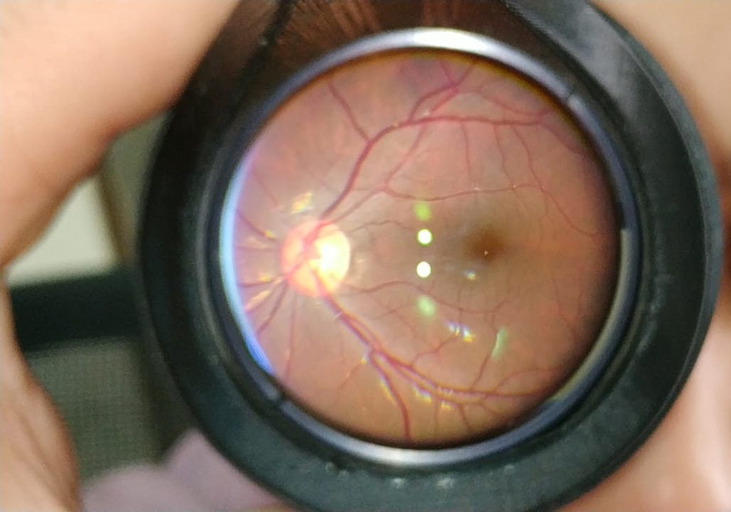
Image Capture: Once properly aligned and focused, the smartphone camera captures digital images and videos of the fundus.
The built-in camera app or specialized software may be used to adjust settings such as exposure and white balance for optimal image quality.
Image Analysis and Sharing: The captured images can be analyzed directly on the smartphone or transmitted to healthcare professionals for interpretation.
Some smartphone apps offer features for basic image processing and analysis, such as image enhancement and annotation.
Benefits of Smartphone Fundus Imaging
The integration of smartphones in fundus examination offers several advantages:
Affordability: Smartphone attachments are significantly cheaper than traditional fundus cameras, making fundus imaging more affordable and accessible, particularly in low-resource settings.
Portability and Accessibility: Smartphones are portable and widely available, allowing for fundus examination to be conducted in various clinical settings, including remote or underserved areas where access to specialized equipment may be limited.
Ease of Use: Smartphone attachments are user-friendly and require minimal training to operate, enabling non-specialist healthcare providers to perform fundus examinations with relative ease.
Patient Engagement: Patients may find smartphone fundus imaging more convenient and less intimidating than traditional methods, potentially leading to increased compliance with screening recommendations.
Telemedicine Applications: The ability to capture and transmit fundus images using smartphones facilitates telemedicine consultations, allowing remote diagnosis and monitoring of ocular conditions by expert clinicians.
Challenges and Considerations
Despite the numerous benefits, smartphone fundus imaging also presents challenges and considerations:
Image Quality and Consistency: Smartphone cameras may not always produce images of the same quality and consistency as traditional fundus cameras, particularly in challenging lighting conditions or with lower-end smartphone models.
Data Security and Privacy: Transmitting patient data, including fundus images, via smartphones raises concerns about data security and patient privacy.
Adequate measures must be implemented to ensure compliance with relevant regulations and standards.
Training and Education: While smartphone fundus imaging is relatively simple to perform, healthcare providers still require training to interpret fundus images accurately and make informed clinical decisions.
Integration with Healthcare Systems: Seamless integration of smartphone fundus imaging into existing healthcare systems may require infrastructure upgrades and workflow modifications to ensure efficient data management and follow-up care.
Conclusion
Fundus examination plays a vital role in the diagnosis and management of various ocular conditions, and fundus cameras are indispensable tools for this purpose.
The integration of smartphones with specialized attachments offers a promising approach to expanding access to fundus imaging, particularly in resource-limited settings.
While smartphone fundus imaging presents challenges, its numerous benefits, including cost-effectiveness, portability, and ease of use, make it a valuable tool for enhancing eye care delivery and promoting early detection and management of ocular diseases.
As technology continues to advance, further research and innovation in smartphone-based fundus imaging are likely to drive improvements in accessibility, affordability, and diagnostic accuracy, ultimately benefiting patients worldwide.